Our Bioinformatics and computational cardiology subgroup is dedicated to advancing the field of medicine, and cardiovascular science in particular, through the integration of cutting-edge computational modeling techniques and state-of-the-art machine learning and bioinformatics suites to provide mechanistic understanding of pathologies and design/evaluation of emerging medical devices. Our team’s expertise in the segmentation, co-registration, and characterization of cardiovascular morphology enables us to create personalized 3D models of vascular beds and organs, which can be used to simulate and analyze the mechanical interactions between medical devices and cardiovascular anatomy. Our patient-specific models allow us to provide clinical insight by predicting clinical events and optimizing therapy for individual patients developing morphology-based heuristics for risk stratification. By translating our computational models and bioinformatics tools to clinical practice, using machine learning algorithms powered by clinical data, we enable the application of precision medicine and personalized therapy for the purpose of treating cardiovascular diseases and developing novel medical devices.
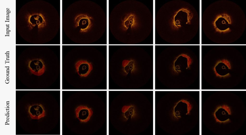
Calcium Analysis: Utilizing machine learning approaches, we can successfully isolate mechanically significant tissues, enabling the three-dimensional evaluation of atherosclerotic structures. This, in turn, permits the prediction of micro-mechanical responses to cardiological treatments, which informs clinical decision-making and contributes to a deeper understanding of disease mechanisms.
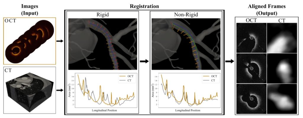
Co-registration Techniques: Advanced image processing and optimization methods enable the integration of multiple coronary imaging modalities into a cohesive dataset. For instance, the trajectory of a virtual catheter within a computed tomography image is optimized to ensure the highest correlation with intravascular imaging data. This image alignment process allows for cross-modal anatomical comparisons and the creation of more accurate computational models of coronary structures.
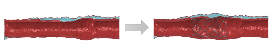
Stenting Simulations: By employing coronary images, we can develop high-precision ‘digital twins’ of individual patients, which serve as the foundation for biomechanical simulations of coronary interventions. These models facilitate the generation of alternative scenarios by modifying patient anatomy, material properties, or intervention parameters, ultimately leading to the optimization of more effective cardiovascular treatments.